How to read dental x-rays
read dental x-rays
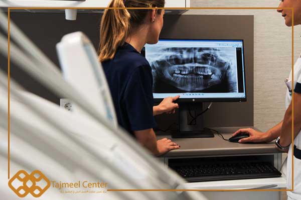
How to read dental x-rays | Our dental x-rays enable us to see what is inside the tooth and what is in the surrounding tissues, so we will provide you with a quick guide on how to read dental x-rays .
What are dental x-rays?
Dental x-rays are images taken with X- rays called X-rays, and what is attached to the jaw bone and the structures and tissues that surround it, which helps the dentist to diagnose the patient’s jaw condition, and x-rays of the mouth may show the tumor, the dental cavity and its structure if it is hidden Such as wisdom teeth, or the basic rules of milk teeth in children, as it shows that bone receding and loss occurred (and the idea of x-rays took shape since it was discovered by the German scientist Rottingen in 1895).
The doctor conducts the x-ray of the tooth when he launches a beam of x-rays that penetrate the tooth structure into the film, then the teeth will appear in a light color and the decay in a dark color because the density of the bone material in them is different.
Types of dental x-rays
The types of dental x-rays are different and the doctor conducts them according to the patient’s therapeutic need, as follows:
- Periapical X-ray: It is a small-sized X-ray, but it looks like 3 or 4 complete teeth.
- Occlusal X-rays: Occlusal X-rays are larger in size than the previous ones, and they fully show the condition of the teeth within the jaw, whether upper or lower; It also shows the surrounding tissues.
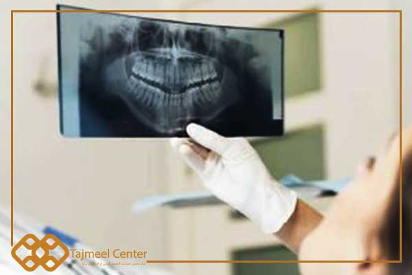
- Panoramic X-rays: Panoramic X-rays show how the teeth and surrounding tissues are in condition, in both the lower and upper jaws, in addition to the jaw joint with the temple. and sinuses.
How to read dental x-rays
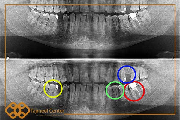
Through the following image, it is clear how to read dental x-rays, which are as follows:
- The area indicated in blue represents an empty space for an extracted tooth.
- The area indicated in red indicates an endodontically treated tooth; The dental canals also appear white due to the presence of a pulp filling in it.
- The area referred to in green indicates the presence of caries within the tooth, as it is gray in the x-ray image, which indicates the presence of caries due to a lack of bone density in it, and that decay and necrosis in the tissues is the reason that enabled the x-rays to pass.
- The area indicated in yellow indicates the amalgam filling, which is white in the picture due to the high density of the mineral so that it does not prevent X-rays from penetrating it.
Dental radiation damage
There are many who fear the harm of dental radiation, including:
- It is known that X-rays cause a change in cells, but this belief can be refuted because the amount of radiation that is used for the tooth is small in a way that is safe for health, as it is equivalent to the rays used in the plane.
- The rays may affect the hard tissue of the tooth, making the metals and adhesives peelable.
- The tooth may be exposed to post-radiation decay.
- Tooth decay and surrounding hard tissues may appear within three months after exposure to radiation.
- Radiation can damage the enamel and crown of the tooth.
In conclusion, the dentist and the laboratory radiologist have sufficient experience in how to read dental x-rays, and anyone can follow this article and learn to read these x-rays easily.
articles that interest you about dental treatment and cosmetic dentistry…..

Dentist In Turkey Prices

Best Dentist Turkey

Best Dental Clinic

Hollywood Smile

Before And After Dental Photos

Dental Reviews

Dental Treatment

Dental Implants

Veneers In Turkey

Dental Crowns

We strive to provide a high level of exceptional service that we expect personally

